July is National Cleft & Craniofacial Awareness & Prevention Month

Story by Jeanie Edgmon
Cleft and craniofacial conditions affect thousands of infants, children, teens, and adults each year. Many are present at birth and represent a diverse group of conditions with innumerable variations. These differences are perhaps among the most sensitive as an individual’s face is one of the most personal features of the body and many can have considerable functional, aesthetic, and social impact within the individual’s life.
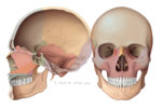
Craniofacial conditions include such things as craniosynostosis, a condition in which the sutures (soft spots) in the skull of an infant close too early, and hemangioma, an abnormally growing blood vessel in the skin that may be present at birth, also known as a port-wine stain, strawberry hemangioma, and salmon patch. Other craniofacial conditions may happen later in life and may be the result of accidents, injuries, and skin diseases.
Craniofacial malformations are involved in three-fourths of all congenital birth defects affecting the head, face, and neck. Of those, cleft lip with or without cleft palate is the most common congenital malformation of the head and the third-most common birth defect, with more than 7,000 born with the condition in the United States each year.
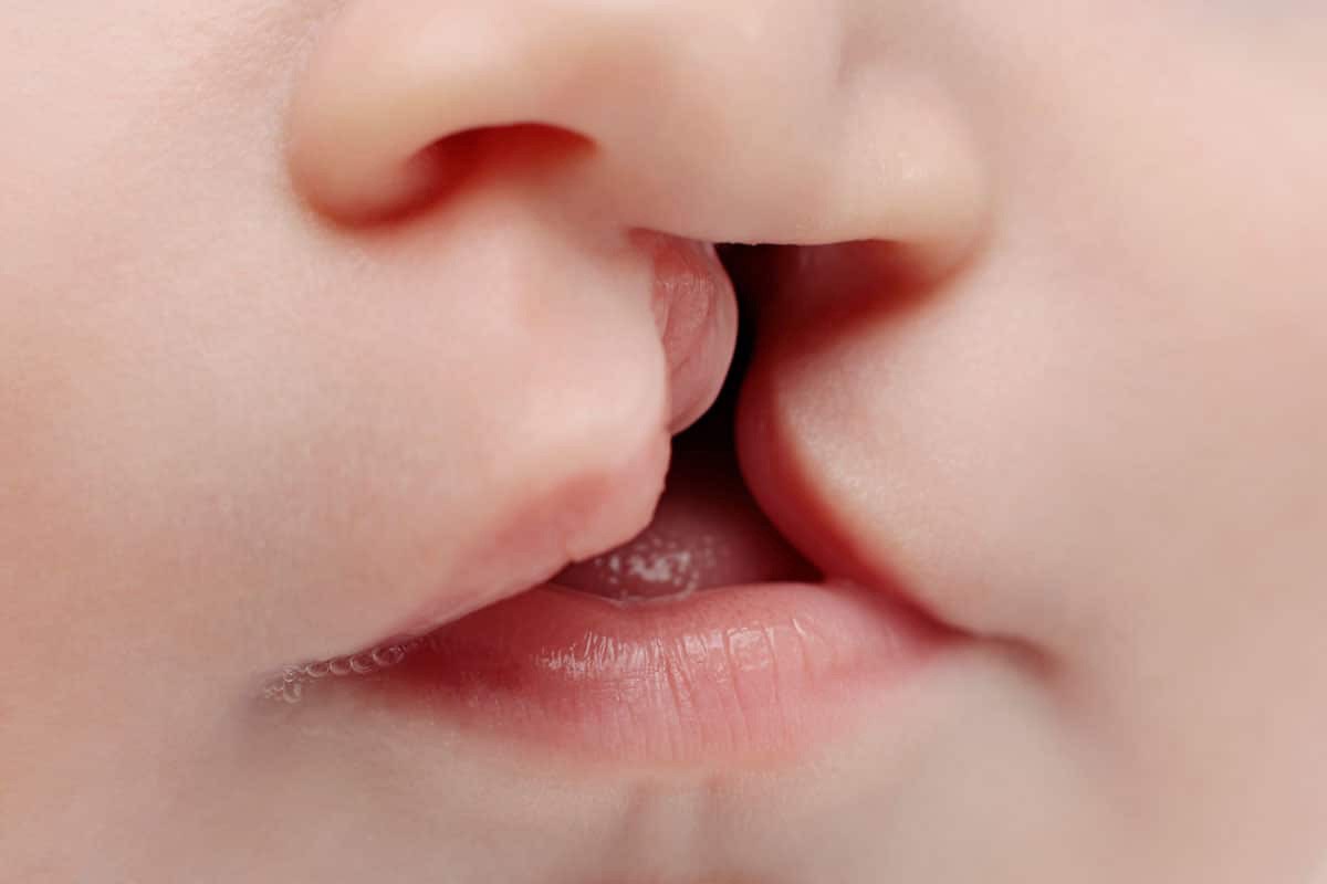
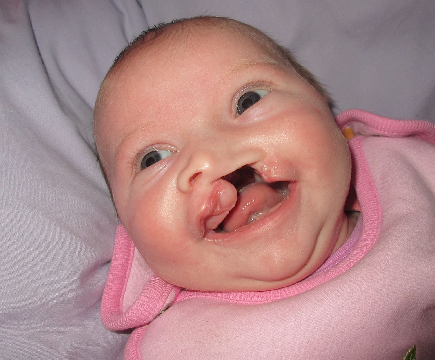
What is a Cleft Lip?
A cleft lip occurs when the tissue that makes up the lip does not join completely before birth. This results in an opening in the upper lip. The opening in the lip can be a small slit or it can be a large opening that goes through the lip into the nose. A cleft lip can be on one or both sides of the lip or in the middle of the lip. According to the Centers for Disease Control and Prevention, about 1 in every 2,800 babies is born with cleft lip without cleft palate in the United States.
What is a Cleft Palate?
The roof of the mouth (palate) is formed between the sixth and ninth weeks of pregnancy. A cleft palate occurs when the tissue that forms the roof of the mouth does not join together completely during pregnancy. In the cleft palate, the front or back of the palate or both may be open. The CDC reports that about 1 in every 1,700 babies is born with cleft palate in the United States.
There are several complications associated with cleft lip and or cleft palate, such as difficulty feeding, ear infections, hearing loss, dental problems, and speech difficulties.
What Causes Cleft and Craniofacial Conditions?
Unfortunately, many things about the cleft are unknown, including the exact cause or causes. Researchers believe that most cases of cleft lip and cleft palate are caused by an interaction of genetic and environmental factors. However, there are several risk factors believed to play a role. These include having a family history of cleft lip or palate; exposure to cigarettes, alcohol, and other substances during pregnancy; or having diabetes or being obese during pregnancy.
Advancing Treatment
The last decade has seen significant progress in the treatment of cleft lip and palate. Technological advances, improved surgical techniques, and a deeper understanding of factors affecting outcomes have all had a serious and positive impact for individuals with cleft and their families.
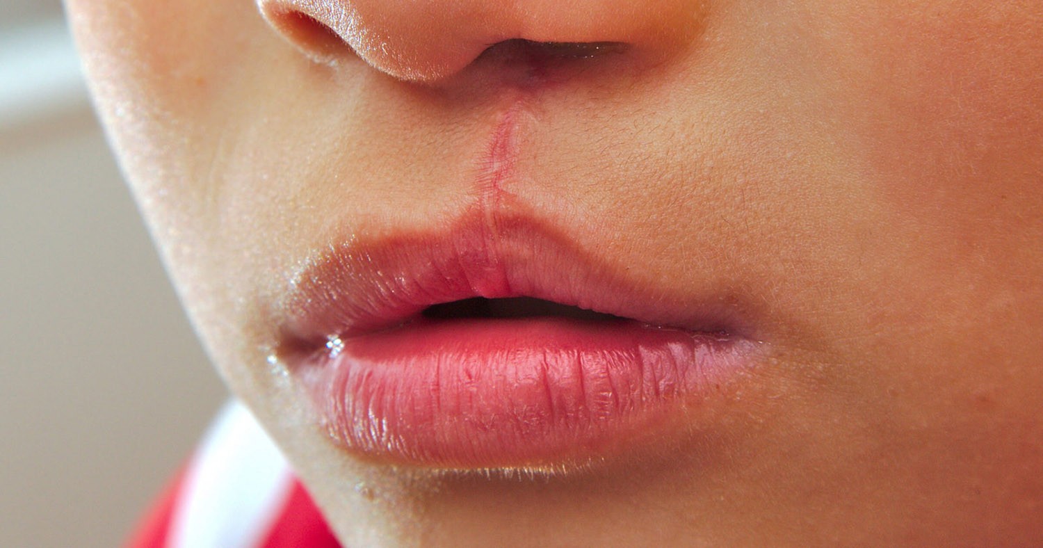
Most significantly, in the past, it would take multiple operations to repair the lip and many more to close the palate. After these surgeries, patients were likely to experience a distinctive speech quality and facial appearance that could cause them to feel stigmatized. Today, the course of treatment and the outcomes are far better. In most cases, a single surgery can reconstruct the lip and nose, and a single surgery can restore the palatal form and function. Both procedures are performed in infancy to normalize the facial structures early, and thus allow the child to progress on a typical course of physical, social, and emotional development. Although revisional surgery is often performed on the lip and nose, these are typically minor and performed later in childhood and adolescence.
One of the factors contributing to better outcomes is advances in imaging. Better imaging allows for an earlier and more accurate diagnosis of a cleft in the prenatal period, improving the parent education process and allowing surgeons to have more information when planning the cleft surgery journey.
Most commonly today, the treatment of cleft and other craniofacial conditions is managed by a multidisciplinary team of specialists over the course of the patient’s development. This team is composed of an array of specialists including nurses, dentists, orthodontists, oral surgeons, otolaryngologists, geneticists, prosthodontists, speech therapists, radiologists, psychologists, feeding specialists, and plastic surgeons. Commonly, the family is integrated as an important part of this team. This approach has significantly improved long-term planning and goals, continuity of care, timing, decision making, execution of high-quality care, and outcomes.
Helping to further these advances are the many teams conducting research in this area. One such institution conducting research is Johns Hopkins whose team is conducting a comparative effectiveness study of two common cleft palate repair techniques: straight-line closure with intra-velar veloplasty (IVVP) and Furlow Z-palatoplasty for children with cleft palate and/or lip (CP±L). Funded by the NIH, this study will analyze speech outcome as primary outcome variables.
While we are making strides in research and treatment, the National Cleft and Craniofacial Awareness Month exists to highlight that there is still significant room for further study to advance our understanding on the origins and treatment of craniofacial disorders.
Despite the large number of children affected by the condition each year, there are still many myths and misconceptions surrounding the condition, the effect it will have, and its treatability.
For More Information on supporting awareness for cleft conditions, their treatment, research, and support, please see www.nccapm.org/faceofchange.html